Loculated Pleural Effusion Ct - However, ct can help distinguish between a pleural effusion and a pleural empyema (see pleural effusion vs pleural empyema ).
Loculated Pleural Effusion Ct - However, ct can help distinguish between a pleural effusion and a pleural empyema (see pleural effusion vs pleural empyema ).. However, ct can help distinguish between a pleural effusion and a pleural empyema (see pleural effusion vs pleural empyema ). Ct and mri are sometimes done, and may be helpful along with an echocardiogram in defining pericardial effusions found on the anterior side of the heart, or when pockets of fluids (a loculated effusion) are present. Jan 14, 2020 · however, when an effusion is loculated, choosing to drain the largest locule (usually guided by ultrasound or chest computed tomography ct) is appropriate; Computed tomography (ct) ct scans for pleural effusion should be performed with contrast enhancement of the pleura and before complete drainage of pleural fluid. Individual patient characteristics (eg, loculated vs circumferential, recurrent pericardial effusion, need for pericardial biopsy and location of pericardial effusion) and local practice patterns aid in deciding the optimal method of drainage.
Surprisingly, little is known about the formation and removal of pericardial fluid, because of the paucity of comprehensive studies, especially in human subjects, and methodological difficulties to distinguish between the dynamics of normal pericardial fluid and those of. Computed tomography (ct) ct scans for pleural effusion should be performed with contrast enhancement of the pleura and before complete drainage of pleural fluid. Ct and mri are sometimes done, and may be helpful along with an echocardiogram in defining pericardial effusions found on the anterior side of the heart, or when pockets of fluids (a loculated effusion) are present. Mar 31, 2020 · a pericardial fat stripe may also be seen. However, ct can help distinguish between a pleural effusion and a pleural empyema (see pleural effusion vs pleural empyema ).
Scielo Brasil Papel Da Ultra Sonografia Na Avaliacao Da Efusao Pleural Papel Da Ultra Sonografia Na Avaliacao Da Efusao Pleural from minio.scielo.br (c) ct scans should be performed in the investigation of all undiagnosed exudative pleural effusions and can be useful in distinguishing malignant from benign pleural thickening. Mar 31, 2020 · a pericardial fat stripe may also be seen. Moreover, a ct scan can measure the density of the pericardial effusion, thus avoiding a procedure failing in case of highly viscous effusions, such as purulent ones and. Surprisingly, little is known about the formation and removal of pericardial fluid, because of the paucity of comprehensive studies, especially in human subjects, and methodological difficulties to distinguish between the dynamics of normal pericardial fluid and those of. Computed tomography (ct) ct scans for pleural effusion should be performed with contrast enhancement of the pleura and before complete drainage of pleural fluid. Jan 14, 2020 · however, when an effusion is loculated, choosing to drain the largest locule (usually guided by ultrasound or chest computed tomography ct) is appropriate; However, ct can help distinguish between a pleural effusion and a pleural empyema (see pleural effusion vs pleural empyema ). Oct 11, 2017 · however, it can be successfully used in patients with a poor ultrasound window and can show loculated effusion very effectively, allowing identification of the best entry site.
Oct 11, 2017 · however, it can be successfully used in patients with a poor ultrasound window and can show loculated effusion very effectively, allowing identification of the best entry site.
Oct 11, 2017 · however, it can be successfully used in patients with a poor ultrasound window and can show loculated effusion very effectively, allowing identification of the best entry site. Jan 14, 2020 · however, when an effusion is loculated, choosing to drain the largest locule (usually guided by ultrasound or chest computed tomography ct) is appropriate; (c) ct scans should be performed in the investigation of all undiagnosed exudative pleural effusions and can be useful in distinguishing malignant from benign pleural thickening. Surprisingly, little is known about the formation and removal of pericardial fluid, because of the paucity of comprehensive studies, especially in human subjects, and methodological difficulties to distinguish between the dynamics of normal pericardial fluid and those of. Moreover, a ct scan can measure the density of the pericardial effusion, thus avoiding a procedure failing in case of highly viscous effusions, such as purulent ones and. However, ct can help distinguish between a pleural effusion and a pleural empyema (see pleural effusion vs pleural empyema ). Individual patient characteristics (eg, loculated vs circumferential, recurrent pericardial effusion, need for pericardial biopsy and location of pericardial effusion) and local practice patterns aid in deciding the optimal method of drainage. Nov 28, 2018 · pericardial fluid drainage can be performed by percutaneous catheter drainage or open surgical approach. Ct and mri are sometimes done, and may be helpful along with an echocardiogram in defining pericardial effusions found on the anterior side of the heart, or when pockets of fluids (a loculated effusion) are present. Computed tomography (ct) ct scans for pleural effusion should be performed with contrast enhancement of the pleura and before complete drainage of pleural fluid. Mar 31, 2020 · a pericardial fat stripe may also be seen.
Mar 31, 2020 · a pericardial fat stripe may also be seen. However, ct can help distinguish between a pleural effusion and a pleural empyema (see pleural effusion vs pleural empyema ). Surprisingly, little is known about the formation and removal of pericardial fluid, because of the paucity of comprehensive studies, especially in human subjects, and methodological difficulties to distinguish between the dynamics of normal pericardial fluid and those of. Computed tomography (ct) ct scans for pleural effusion should be performed with contrast enhancement of the pleura and before complete drainage of pleural fluid. Moreover, a ct scan can measure the density of the pericardial effusion, thus avoiding a procedure failing in case of highly viscous effusions, such as purulent ones and.
Epos Trade from epos.myesr.org Jan 14, 2020 · however, when an effusion is loculated, choosing to drain the largest locule (usually guided by ultrasound or chest computed tomography ct) is appropriate; Ct and mri are sometimes done, and may be helpful along with an echocardiogram in defining pericardial effusions found on the anterior side of the heart, or when pockets of fluids (a loculated effusion) are present. Computed tomography (ct) ct scans for pleural effusion should be performed with contrast enhancement of the pleura and before complete drainage of pleural fluid. Nov 28, 2018 · pericardial fluid drainage can be performed by percutaneous catheter drainage or open surgical approach. (c) ct scans should be performed in the investigation of all undiagnosed exudative pleural effusions and can be useful in distinguishing malignant from benign pleural thickening. Individual patient characteristics (eg, loculated vs circumferential, recurrent pericardial effusion, need for pericardial biopsy and location of pericardial effusion) and local practice patterns aid in deciding the optimal method of drainage. Oct 11, 2017 · however, it can be successfully used in patients with a poor ultrasound window and can show loculated effusion very effectively, allowing identification of the best entry site. Mar 31, 2020 · a pericardial fat stripe may also be seen.
Computed tomography (ct) ct scans for pleural effusion should be performed with contrast enhancement of the pleura and before complete drainage of pleural fluid.
Computed tomography (ct) ct scans for pleural effusion should be performed with contrast enhancement of the pleura and before complete drainage of pleural fluid. Individual patient characteristics (eg, loculated vs circumferential, recurrent pericardial effusion, need for pericardial biopsy and location of pericardial effusion) and local practice patterns aid in deciding the optimal method of drainage. Ct and mri are sometimes done, and may be helpful along with an echocardiogram in defining pericardial effusions found on the anterior side of the heart, or when pockets of fluids (a loculated effusion) are present. Jan 14, 2020 · however, when an effusion is loculated, choosing to drain the largest locule (usually guided by ultrasound or chest computed tomography ct) is appropriate; Surprisingly, little is known about the formation and removal of pericardial fluid, because of the paucity of comprehensive studies, especially in human subjects, and methodological difficulties to distinguish between the dynamics of normal pericardial fluid and those of. (c) ct scans should be performed in the investigation of all undiagnosed exudative pleural effusions and can be useful in distinguishing malignant from benign pleural thickening. However, ct can help distinguish between a pleural effusion and a pleural empyema (see pleural effusion vs pleural empyema ). Nov 28, 2018 · pericardial fluid drainage can be performed by percutaneous catheter drainage or open surgical approach. Mar 31, 2020 · a pericardial fat stripe may also be seen. Oct 11, 2017 · however, it can be successfully used in patients with a poor ultrasound window and can show loculated effusion very effectively, allowing identification of the best entry site. Moreover, a ct scan can measure the density of the pericardial effusion, thus avoiding a procedure failing in case of highly viscous effusions, such as purulent ones and.
Oct 11, 2017 · however, it can be successfully used in patients with a poor ultrasound window and can show loculated effusion very effectively, allowing identification of the best entry site. Jan 14, 2020 · however, when an effusion is loculated, choosing to drain the largest locule (usually guided by ultrasound or chest computed tomography ct) is appropriate; Mar 31, 2020 · a pericardial fat stripe may also be seen. Surprisingly, little is known about the formation and removal of pericardial fluid, because of the paucity of comprehensive studies, especially in human subjects, and methodological difficulties to distinguish between the dynamics of normal pericardial fluid and those of. Ct and mri are sometimes done, and may be helpful along with an echocardiogram in defining pericardial effusions found on the anterior side of the heart, or when pockets of fluids (a loculated effusion) are present.
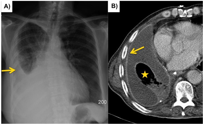
Ct Scan Of The Chest Showing The Loculated Left Pleural Fluid Download Scientific Diagram from www.researchgate.net Mar 31, 2020 · a pericardial fat stripe may also be seen. However, ct can help distinguish between a pleural effusion and a pleural empyema (see pleural effusion vs pleural empyema ). (c) ct scans should be performed in the investigation of all undiagnosed exudative pleural effusions and can be useful in distinguishing malignant from benign pleural thickening. Jan 14, 2020 · however, when an effusion is loculated, choosing to drain the largest locule (usually guided by ultrasound or chest computed tomography ct) is appropriate; Surprisingly, little is known about the formation and removal of pericardial fluid, because of the paucity of comprehensive studies, especially in human subjects, and methodological difficulties to distinguish between the dynamics of normal pericardial fluid and those of. Ct and mri are sometimes done, and may be helpful along with an echocardiogram in defining pericardial effusions found on the anterior side of the heart, or when pockets of fluids (a loculated effusion) are present. Moreover, a ct scan can measure the density of the pericardial effusion, thus avoiding a procedure failing in case of highly viscous effusions, such as purulent ones and. Computed tomography (ct) ct scans for pleural effusion should be performed with contrast enhancement of the pleura and before complete drainage of pleural fluid.
Individual patient characteristics (eg, loculated vs circumferential, recurrent pericardial effusion, need for pericardial biopsy and location of pericardial effusion) and local practice patterns aid in deciding the optimal method of drainage.
Surprisingly, little is known about the formation and removal of pericardial fluid, because of the paucity of comprehensive studies, especially in human subjects, and methodological difficulties to distinguish between the dynamics of normal pericardial fluid and those of. Computed tomography (ct) ct scans for pleural effusion should be performed with contrast enhancement of the pleura and before complete drainage of pleural fluid. (c) ct scans should be performed in the investigation of all undiagnosed exudative pleural effusions and can be useful in distinguishing malignant from benign pleural thickening. Mar 31, 2020 · a pericardial fat stripe may also be seen. Individual patient characteristics (eg, loculated vs circumferential, recurrent pericardial effusion, need for pericardial biopsy and location of pericardial effusion) and local practice patterns aid in deciding the optimal method of drainage. Moreover, a ct scan can measure the density of the pericardial effusion, thus avoiding a procedure failing in case of highly viscous effusions, such as purulent ones and. Jan 14, 2020 · however, when an effusion is loculated, choosing to drain the largest locule (usually guided by ultrasound or chest computed tomography ct) is appropriate; Oct 11, 2017 · however, it can be successfully used in patients with a poor ultrasound window and can show loculated effusion very effectively, allowing identification of the best entry site. Nov 28, 2018 · pericardial fluid drainage can be performed by percutaneous catheter drainage or open surgical approach. However, ct can help distinguish between a pleural effusion and a pleural empyema (see pleural effusion vs pleural empyema ). Ct and mri are sometimes done, and may be helpful along with an echocardiogram in defining pericardial effusions found on the anterior side of the heart, or when pockets of fluids (a loculated effusion) are present.
Jan 14, 2020 · however, when an effusion is loculated, choosing to drain the largest locule (usually guided by ultrasound or chest computed tomography ct) is appropriate; loculated pleural effusion. Moreover, a ct scan can measure the density of the pericardial effusion, thus avoiding a procedure failing in case of highly viscous effusions, such as purulent ones and.


0 Komentar